

What Do Animal Cells Have The Plant Cells Don't
Developmental biology is the study of the procedure past which animals and plants abound and develop. Developmental biology likewise encompasses the biological science of regeneration, asexual reproduction, metamorphosis, and the growth and differentiation of stem cells in the developed organism.
Perspectives [edit]
The main processes involved in the embryonic development of animals are: tissue patterning (via regional specification and patterned prison cell differentiation); tissue growth; and tissue morphogenesis.
- Regional specification refers to the processes that create spatial pattern in a brawl or sheet of initially similar cells. This generally involves the activity of cytoplasmic determinants, located inside parts of the fertilized egg, and of inductive signals emitted from signaling centers in the embryo. The early on stages of regional specification do not generate functional differentiated cells, but cell populations committed to develop to a specific region or part of the organism. These are divers past the expression of specific combinations of transcription factors.
- Cell differentiation relates specifically to the formation of functional cell types such every bit nerve, muscle, secretory epithelia etc. Differentiated cells comprise large amounts of specific proteins associated with the cell role.
- Morphogenesis relates to the formation of 3-dimensional shape. Information technology mainly involves the orchestrated movements of prison cell sheets and of individual cells. Morphogenesis is important for creating the three germ layers of the early embryo (ectoderm, mesoderm and endoderm) and for edifice upwards complex structures during organ evolution.
- Tissue growth involves both an overall increase in tissue size, and as well the differential growth of parts (allometry) which contributes to morphogenesis. Growth more often than not occurs through prison cell proliferation but besides through changes of jail cell size or the degradation of extracellular materials.
The development of plants involves similar processes to that of animals. Yet plant cells are mostly immotile so morphogenesis is achieved by differential growth, without prison cell movements. Likewise, the inductive signals and the genes involved are different from those that command fauna development.
Developmental processes [edit]
Cell differentiation [edit]
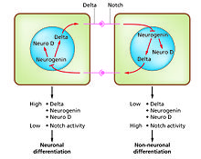
The Notch-delta organization in neurogenesis.(Slack Essential Dev Biol Fig 14.12a)
Prison cell differentiation is the process whereby different functional jail cell types arise in evolution. For example, neurons, musculus fibers and hepatocytes (liver cells) are well known types of differentiated cells. Differentiated cells usually produce large amounts of a few proteins that are required for their specific function and this gives them the characteristic advent that enables them to be recognized under the low-cal microscope. The genes encoding these proteins are highly active. Typically their chromatin structure is very open, assuasive access for the transcription enzymes, and specific transcription factors demark to regulatory sequences in the DNA in guild to actuate factor expression.[one] [2] For instance, NeuroD is a key transcription factor for neuronal differentiation, myogenin for muscle differentiation, and HNF4 for hepatocyte differentiation. Prison cell differentiation is usually the final stage of development, preceded by several states of delivery which are not visibly differentiated. A unmarried tissue, formed from a single type of progenitor cell or stalk prison cell, often consists of several differentiated cell types. Control of their germination involves a procedure of lateral inhibition,[3] based on the backdrop of the Notch signaling pathway.[iv] For example, in the neural plate of the embryo this organisation operates to generate a population of neuronal precursor cells in which NeuroD is highly expressed.
Regeneration [edit]
Regeneration indicates the power to regrow a missing part.[v] This is very prevalent among plants, which show continuous growth, and also among colonial animals such as hydroids and ascidians. But most interest by developmental biologists has been shown in the regeneration of parts in free living animals. In particular four models have been the subject area of much investigation. Ii of these have the ability to regenerate whole bodies: Hydra, which can regenerate any function of the polyp from a small fragment,[six] and planarian worms, which tin can ordinarily regenerate both heads and tails.[vii] Both of these examples have continuous prison cell turnover fed by stalk cells and, at least in planaria, at least some of the stem cells have been shown to exist pluripotent.[8] The other two models show simply distal regeneration of appendages. These are the insect appendages, unremarkably the legs of hemimetabolous insects such equally the cricket,[9] and the limbs of urodele amphibians.[x] Considerable data is now available well-nigh amphibian limb regeneration and information technology is known that each jail cell blazon regenerates itself, except for connective tissues where at that place is considerable interconversion between cartilage, dermis and tendons. In terms of the blueprint of structures, this is controlled by a re-activation of signals active in the embryo. There is still argue about the old question of whether regeneration is a "pristine" or an "adaptive" holding.[11] If the former is the case, with improved knowledge, we might expect to exist able to improve regenerative power in humans. If the latter, then each instance of regeneration is presumed to take arisen by natural selection in circumstances particular to the species, so no general rules would exist expected.
Embryonic development of animals [edit]
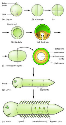
Generalized scheme of embryonic evolution. Slack "Essential Developmental Biology" Fig.ii.eight

The sperm and egg fuse in the process of fertilization to form a fertilized egg, or zygote.[12] This undergoes a menses of divisions to form a ball or sheet of like cells called a blastula or blastoderm. These cell divisions are usually rapid with no growth and then the daughter cells are half the size of the mother cell and the whole embryo stays about the same size. They are chosen cleavage divisions.
Mouse epiblast primordial germ cells (come across Effigy: "The initial stages of human embryogenesis") undergo extensive epigenetic reprogramming.[xiii] This process involves genome-wide Deoxyribonucleic acid demethylation, chromatin reorganization and epigenetic imprint erasure leading to totipotency.[13] DNA demethylation is carried out by a process that utilizes the DNA base of operations excision repair pathway.[14]
Morphogenetic movements catechumen the prison cell mass into a three layered structure consisting of multicellular sheets called ectoderm, mesoderm and endoderm. These sheets are known as germ layers. This is the process of gastrulation. During cleavage and gastrulation the first regional specification events occur. In improver to the formation of the three germ layers themselves, these often generate extraembryonic structures, such as the mammalian placenta, needed for support and nutrition of the embryo,[15] and also establish differences of commitment along the anteroposterior axis (head, body and tail).[16]
Regional specification is initiated by the presence of cytoplasmic determinants in i part of the zygote. The cells that contain the determinant become a signaling center and emit an inducing factor. Because the inducing cistron is produced in one identify, diffuses away, and decays, information technology forms a concentration gradient, loftier near the source cells and low further abroad.[17] [18] The remaining cells of the embryo, which practise not contain the determinant, are competent to respond to dissimilar concentrations by upregulating specific developmental command genes. This results in a series of zones becoming fix, arranged at progressively greater distance from the signaling heart. In each zone a different combination of developmental command genes is upregulated.[19] These genes encode transcription factors which upregulate new combinations of gene activity in each region. Among other functions, these transcription factors control expression of genes conferring specific adhesive and movement properties on the cells in which they are active. Because of these unlike morphogenetic properties, the cells of each germ layer motility to form sheets such that the ectoderm ends up on the exterior, mesoderm in the middle, and endoderm on the inside.[twenty] [21] Morphogenetic movements not simply change the shape and construction of the embryo, but past bringing cell sheets into new spatial relationships they also make possible new phases of signaling and response between them.
Growth in embryos is mostly autonomous.[22] For each territory of cells the growth charge per unit is controlled by the combination of genes that are active. Free-living embryos do not grow in mass as they take no external food supply. But embryos fed by a placenta or extraembryonic yolk supply can grow very fast, and changes to relative growth charge per unit between parts in these organisms aid to produce the final overall anatomy.
The whole process needs to exist coordinated in time and how this is controlled is not understood. In that location may exist a master clock able to communicate with all parts of the embryo that controls the class of events, or timing may depend simply on local causal sequences of events.[23]
Metamorphosis [edit]
Developmental processes are very evident during the process of metamorphosis. This occurs in various types of animal. Well-known examples are seen in frogs, which commonly hatch as a tadpole and metamorphoses to an developed frog, and certain insects which hatch as a larva and then become remodeled to the developed form during a pupal stage.
All the developmental processes listed to a higher place occur during metamorphosis. Examples that have been especially well studied include tail loss and other changes in the polliwog of the frog Xenopus,[24] [25] and the biology of the imaginal discs, which generate the adult body parts of the fly Drosophila melanogaster.[26] [27]
Found evolution [edit]
Plant development is the process by which structures originate and mature as a plant grows. Information technology is studied in found beefcake and plant physiology as well as found morphology.
Plants constantly produce new tissues and structures throughout their life from meristems[28] located at the tips of organs, or betwixt mature tissues. Thus, a living plant e'er has embryonic tissues. By dissimilarity, an animal embryo will very early produce all of the body parts that it will ever have in its life. When the brute is born (or hatches from its egg), information technology has all its torso parts and from that point will only abound larger and more mature.
The backdrop of organization seen in a plant are emergent properties which are more than the sum of the individual parts. "The associates of these tissues and functions into an integrated multicellular organism yields non only the characteristics of the carve up parts and processes just also quite a new prepare of characteristics which would not have been predictable on the ground of examination of the separate parts."[29]
Growth [edit]
A vascular plant begins from a single celled zygote, formed past fertilisation of an egg cell by a sperm cell. From that point, it begins to split to form a plant embryo through the process of embryogenesis. As this happens, the resulting cells will organize so that 1 stop becomes the first root, while the other end forms the tip of the shoot. In seed plants, the embryo will develop one or more "seed leaves" (cotyledons). By the stop of embryogenesis, the young plant volition have all the parts necessary to begin its life.
One time the embryo germinates from its seed or parent plant, it begins to produce additional organs (leaves, stems, and roots) through the procedure of organogenesis. New roots abound from root meristems located at the tip of the root, and new stems and leaves grow from shoot meristems located at the tip of the shoot.[30] Branching occurs when small clumps of cells left behind by the meristem, and which have not notwithstanding undergone cellular differentiation to form a specialized tissue, begin to grow every bit the tip of a new root or shoot. Growth from whatever such meristem at the tip of a root or shoot is termed primary growth and results in the lengthening of that root or shoot. Secondary growth results in widening of a root or shoot from divisions of cells in a cambium.[31]
In addition to growth by cell division, a plant may grow through cell elongation.[32] This occurs when individual cells or groups of cells grow longer. Not all plant cells volition abound to the same length. When cells on 1 side of a stem grow longer and faster than cells on the other side, the stem will bend to the side of the slower growing cells equally a result. This directional growth can occur via a plant'due south response to a particular stimulus, such as low-cal (phototropism), gravity (gravitropism), h2o, (hydrotropism), and concrete contact (thigmotropism).
Found growth and development are mediated by specific establish hormones and plant growth regulators (PGRs) (Ross et al. 1983).[33] Endogenous hormone levels are influenced by plant historic period, common cold hardiness, dormancy, and other metabolic conditions; photoperiod, drought, temperature, and other external environmental conditions; and exogenous sources of PGRs, e.yard., externally applied and of rhizospheric origin.
Morphological variation [edit]
Plants exhibit natural variation in their form and construction. While all organisms vary from individual to individual, plants exhibit an additional blazon of variation. Within a single individual, parts are repeated which may differ in grade and structure from other similar parts. This variation is well-nigh easily seen in the leaves of a plant, though other organs such every bit stems and flowers may show similar variation. There are three chief causes of this variation: positional effects, environmental effects, and juvenility.
Evolution of constitute morphology [edit]
Transcription factors and transcriptional regulatory networks play key roles in plant morphogenesis and their development. During plant landing, many novel transcription factor families emerged and are preferentially wired into the networks of multicellular development, reproduction, and organ development, contributing to more than complex morphogenesis of land plants.[34]
Most land plants share a common ancestor, multicellular algae. An example of the evolution of plant morphology is seen in charophytes. Studies have shown that charophytes take traits that are homologous to land plants. At that place are two master theories of the development of plant morphology, these theories are the homologous theory and the antithetic theory. The commonly accepted theory for the evolution of plant morphology is the antithetic theory. The antithetic theory states that the multiple mitotic divisions that take place before meiosis, cause the development of the sporophyte. Then the sporophyte will evolution as an independent organism.[35]
Developmental model organisms [edit]
Much of developmental biology enquiry in contempo decades has focused on the apply of a modest number of model organisms. It has turned out that at that place is much conservation of developmental mechanisms across the animal kingdom. In early development different vertebrate species all use essentially the same anterior signals and the same genes encoding regional identity. Even invertebrates utilize a similar repertoire of signals and genes although the body parts formed are significantly dissimilar. Model organisms each take some detail experimental advantages which accept enabled them to become popular among researchers. In one sense they are "models" for the whole animal kingdom, and in another sense they are "models" for man development, which is hard to study directly for both ethical and practical reasons. Model organisms accept been most useful for elucidating the broad nature of developmental mechanisms. The more detail is sought, the more they differ from each other and from humans.
Plants [edit]
- Thale cress (Arabidopsis thaliana)[36]
Vertebrates [edit]
- Frog: Xenopus [36] (X. laevis and X. tropicalis).[37] [38] Good embryo supply. Especially suitable for microsurgery.
- Zebrafish: Danio rerio.[39] Skillful embryo supply. Well developed genetics.
- Chicken: Gallus gallus.[40] Early stages similar to mammal, merely microsurgery easier. Depression toll.
- Mouse: Mus musculus.[41] A mammal[36] with well developed genetics.
Invertebrates [edit]
- Fruit fly: Drosophila melanogaster.[42] Good embryo supply. Well adult genetics.
- Nematode: Caenorhabditis elegans.[43] Adept embryo supply. Well adult genetics. Low cost.
Unicellular [edit]
- Algae: Chlamydomonas [36]
- Yeast: Saccharomyces [36]
Others [edit]
Besides popular for some purposes take been sea urchins[44] [36] and ascidians.[45] For studies of regeneration urodele amphibians such as the axolotl Ambystoma mexicanum are used,[46] and too planarian worms such every bit Schmidtea mediterranea.[seven] Organoids have also been demonstrated as an efficient model for development.[47] Plant development has focused on the thale cress Arabidopsis thaliana as a model organism.[48]...
See also [edit]
- Blastocyst
- Body plan
- Cell signaling
- Cell signaling networks
- Embryology
- Enhancer
- Fish development
- Factor regulatory network
- Ontogenesis
- Found evolutionary developmental biology
- Promoter (biology)
- Signal transduction
- Teratology
References [edit]
- ^ Li B, Carey K, Workman JL (February 2007). "The office of chromatin during transcription". Jail cell. 128 (4): 707–19. doi:10.1016/j.cell.2007.01.015. PMID 17320508.
- ^ Heintzman ND, Stuart RK, Hon G, Fu Y, Ching CW, Hawkins RD, et al. (March 2007). "Distinct and predictive chromatin signatures of transcriptional promoters and enhancers in the human being genome". Nature Genetics. 39 (iii): 311–8. doi:10.1038/ng1966. PMID 17277777. S2CID 1595885.
- ^ Meinhardt H, Gierer A (2000). "Pattern germination by local self-activation and lateral inhibition" (PDF). BioEssays. 22 (8): 753–760. CiteSeerX10.1.one.477.439. doi:10.1002/1521-1878(200008)22:viii<753::assistance-bies9>3.0.co;2-z. PMID 10918306. Archived (PDF) from the original on 2017-x-27.
- ^ Sprinzak D, Lakhanpal A, Lebon L, Santat LA, Fontes ME, Anderson GA, et al. (May 2010). "Cis-interactions between Notch and Delta generate mutually exclusive signalling states". Nature. 465 (7294): 86–90. Bibcode:2010Natur.465...86S. doi:ten.1038/nature08959. PMC2886601. PMID 20418862.
- ^ Carlson BM (2007). Principles of Regenerative Biology. Burlington MA: Bookish Press.
- ^ Bosch TC (March 2007). "Why polyps regenerate and we don't: towards a cellular and molecular framework for Hydra regeneration". Developmental Biology. 303 (two): 421–33. doi:10.1016/j.ydbio.2006.12.012. PMID 17234176.
- ^ a b Reddien PW, Sánchez Alvarado A (2004). "Fundamentals of planarian regeneration". Annual Review of Cell and Developmental Biological science. 20: 725–57. doi:x.1146/annurev.cellbio.20.010403.095114. PMID 15473858. S2CID 1320382.
- ^ Wagner DE, Wang IE, Reddien PW (May 2011). "Clonogenic neoblasts are pluripotent adult stem cells that underlie planarian regeneration". Science. 332 (6031): 811–6. Bibcode:2011Sci...332..811W. doi:10.1126/scientific discipline.1203983. PMC3338249. PMID 21566185.
- ^ Nakamura T, Mito T, Bando T, Ohuchi H, Noji Due south (Jan 2008). "Dissecting insect leg regeneration through RNA interference". Cellular and Molecular Life Sciences. 65 (1): 64–72. doi:x.1007/s00018-007-7432-0. PMID 18030418.
- ^ Simon A, Tanaka EM (2013). "Limb regeneration". Wiley Interdisciplinary Reviews. Developmental Biology. ii (2): 291–300. doi:x.1002/wdev.73. PMID 24009038. S2CID 13158705.
- ^ Slack JM (2013). "Chapter 20". Essential Developmental Biology. Oxford: Wiley-Blackwell.
- ^ Jungnickel MK, Sutton KA, Florman HM (August 2003). "In the beginning: lessons from fertilization in mice and worms". Jail cell. 114 (iv): 401–4. doi:10.1016/s0092-8674(03)00648-two. PMID 12941269.
- ^ a b Hackett JA, Sengupta R, Zylicz JJ, Murakami K, Lee C, Downward TA, Surani MA (Jan 2013). "Germline DNA demethylation dynamics and imprint erasure through 5-hydroxymethylcytosine". Science. 339 (6118): 448–52. Bibcode:2013Sci...339..448H. doi:10.1126/science.1229277. PMC3847602. PMID 23223451.
- ^ Hajkova P, Jeffries SJ, Lee C, Miller North, Jackson SP, Surani MA (July 2010). "Genome-broad reprogramming in the mouse germ line entails the base of operations excision repair pathway". Science. 329 (5987): 78–82. Bibcode:2010Sci...329...78H. doi:x.1126/scientific discipline.1187945. PMC3863715. PMID 20595612.
- ^ Steven DH, ed. (1975). Comparative Placentation. London: Academic Press.
- ^ Kimelman D, Martin BL (2012). "Anterior-posterior patterning in early development: 3 strategies". Wiley Interdisciplinary Reviews. Developmental Biological science. i (two): 253–66. doi:10.1002/wdev.25. PMC5560123. PMID 23801439.
- ^ Slack JM (1987). "Morphogenetic gradients - by and present". Trends in Biochemical Sciences. 12: 200–204. doi:x.1016/0968-0004(87)90094-six.
- ^
- ^ Dahmann C, Oates AC, Make M (Jan 2011). "Boundary formation and maintenance in tissue development". Nature Reviews. Genetics. 12 (1): 43–55. doi:10.1038/nrg2902. PMID 21164524. S2CID 1805261.
- ^ Hardin J, Walston T (August 2004). "Models of morphogenesis: the mechanisms and mechanics of prison cell rearrangement". Current Opinion in Genetics & Development. 14 (4): 399–406. doi:10.1016/j.gde.2004.06.008. PMID 15261656.
- ^ Hammerschmidt Thou, Wedlich D (Nov 2008). "Regulated adhesion as a driving force of gastrulation movements". Development. 135 (22): 3625–41. doi:10.1242/dev.015701. PMID 18952908.
- ^ O'Farrell PH (2003). "How metazoans reach their full size: the natural history of bigness.". In Hall MN, Raff M, Thomas Chiliad (eds.). Cell Growth: Control of Cell Size. Cold Leap Harbor Laboratory Printing. pp. 1–21.
- ^ Moss EG, Romer-Seibert J (2014). "Prison cell-intrinsic timing in brute evolution". Wiley Interdisciplinary Reviews. Developmental Biology. iii (five): 365–77. doi:10.1002/wdev.145. PMID 25124757. S2CID 29029979.
- ^ Tata JR (1996). "Amphibian metamorphosis: an exquisite model for hormonal regulation of postembryonic development in vertebrates". Development, Growth and Differentiation. 38 (three): 223–231. doi:10.1046/j.1440-169x.1996.t01-two-00001.x. S2CID 84081060.
- ^ Brown DD, Cai 50 (June 2007). "Amphibian metamorphosis". Developmental Biology. 306 (i): xx–33. doi:ten.1016/j.ydbio.2007.03.021. PMC1945045. PMID 17449026.
- ^ Cohen SM (1993). "Imaginal Disc Development.". In Bate Thou, Martinez-Arias M (eds.). The Development of Drosophila melanogaster. Cold Spring Harbor Press.
- ^ Maves L, Schubiger G (Oct 2003). "Transdetermination in Drosophila imaginal discs: a model for understanding pluripotency and selector cistron maintenance". Current Opinion in Genetics & Development. 13 (5): 472–ix. doi:10.1016/j.gde.2003.08.006. PMID 14550411.
- ^ Bäurle I, Laux T (October 2003). "Apical meristems: the constitute'due south fountain of youth". Review. BioEssays. 25 (ten): 961–lxx. doi:10.1002/bies.10341. PMID 14505363.
- ^ Leopold Ac (1964). Constitute Growth and Evolution . New York: McGraw-Colina. p. 183.
- ^ Brand U, Hobe Grand, Simon R (February 2001). "Functional domains in plant shoot meristems". Review. BioEssays. 23 (2): 134–41. doi:10.1002/1521-1878(200102)23:ii<134::Aid-BIES1020>3.0.CO;ii-3. PMID 11169586.
- ^ Barlow P (May 2005). "Patterned cell decision in a plant tissue: the secondary phloem of trees". BioEssays. 27 (5): 533–41. doi:x.1002/bies.20214. PMID 15832381.
- ^ Pacifici E, Di Mambro R, Dello Ioio R, Costantino P, Sabatini S (August 2018). "Arabidopsis root". The EMBO Journal. 37 (sixteen). doi:ten.15252/embj.201899134. PMC6092616. PMID 30012836.
- ^ Ross SD, Pharis RP, Binder WD (1983). "Growth regulators and conifers: their physiology and potential uses in forestry.". In Nickell LG (ed.). Found growth regulating chemicals. Vol. 2. Boca Raton, FL: CRC Press. pp. 35–78.
- ^ Jin J, He K, Tang X, Li Z, Lv L, Zhao Y, et al. (July 2015). "An Arabidopsis Transcriptional Regulatory Map Reveals Distinct Functional and Evolutionary Features of Novel Transcription Factors". Molecular Biology and Evolution. 32 (seven): 1767–73. doi:10.1093/molbev/msv058. PMC4476157. PMID 25750178. Archived from the original on 2016-06-02.
- ^ Pires, Nuno D.; Dolan, Liam (2012-02-xix). "Morphological evolution in land plants: new designs with old genes". Philosophical Transactions of the Royal Society B: Biological Sciences. 367 (1588): 508–518. doi:ten.1098/rstb.2011.0252. ISSN 0962-8436. PMC3248709. PMID 22232763.
- ^ a b c d due east f Friedman, William E. (1999). "Expression of the cell cycle in sperm of Arabidopsis: implications for understanding patterns of gametogenesis and fertilization in plants and other eukaryotes". Evolution. The Company of Biologists. 126 (5): 1065–75. doi:10.1242/dev.126.5.1065. ISSN 0950-1991. PMID 9927606. S2CID 13397345.
- ^ Nieuwkoop PD, Faber J (1967). Normal tabular array of Xenopus laevis (Daudin). Due north-The netherlands, Amsterdam.
- ^ Harland RM, Grainger RM (December 2011). "Xenopus research: metamorphosed by genetics and genomics". Trends in Genetics. 27 (12): 507–fifteen. doi:10.1016/j.tig.2011.08.003. PMC3601910. PMID 21963197.
- ^ Lawson ND, Wolfe SA (July 2011). "Forward and reverse genetic approaches for the assay of vertebrate development in the zebrafish". Developmental Cell. 21 (ane): 48–64. doi:x.1016/j.devcel.2011.06.007. PMID 21763608.
- ^ Rashidi H, Sottile V (Apr 2009). "The chick embryo: hatching a model for contemporary biomedical research". BioEssays. 31 (iv): 459–65. doi:10.1002/bies.200800168. PMID 19274658. S2CID 5489431.
- ^ Behringer R, Gertsenstein Chiliad, Vintersten 1000, Nagy Grand (2014). Manipulating the Mouse Embryo. A Laboratory Manual (Fourth ed.). Cold Spring Harbor, NY: Cold Spring Harbor Laboratory Printing.
- ^ St Johnston D (March 2002). "The fine art and design of genetic screens: Drosophila melanogaster". Nature Reviews. Genetics. 3 (3): 176–88. doi:10.1038/nrg751. PMID 11972155. S2CID 195368351.
- ^ Riddle DL, Blumenthal T, Meyer BJ, Priess JR (1997). C.elegans Two. Cold Spring Harbor, NY: Cold Spring Harbor Laboratory Printing.
- ^ Ettensohn CA, Sugariness HC (2000). "Patterning the early sea urchin embryo". Current Topics in Developmental Biology Volume l. Curr. Superlative. Dev. Biol. Current Topics in Developmental Biology. Vol. 50. Academic Press. pp. ane–44. doi:10.1016/S0070-2153(00)50002-seven. ISBN9780121531508. PMID 10948448.
- ^ Lemaire P (June 2011). "Evolutionary crossroads in developmental biology: the tunicates". Development. 138 (11): 2143–52. doi:10.1242/dev.048975. PMID 21558365.
- ^ Nacu Eastward, Tanaka EM (2011). "Limb regeneration: a new development?". Annual Review of Prison cell and Developmental Biology. 27: 409–forty. doi:10.1146/annurev-cellbio-092910-154115. PMID 21801016.
- ^ Ader M, Tanaka EM (Dec 2014). "Modeling man development in 3D culture". Electric current Opinion in Cell Biology. 31: 23–8. doi:10.1016/j.ceb.2014.06.013. PMID 25033469.
- ^ Weigel D, Glazebrook J (2002). Arabidopsis. A Laboratory Manual. Cold Jump Harbor, NY: Cold Spring Harbor Laboratory Press.
Further reading [edit]
- Gilbert SF (2013). Developmental Biology. Sunderland, Mass.: Sinauer Associates Inc.
- Slack JM (2013). Essential Developmental Biology. Oxford: Wiley-Blackwell.
- Wolpert L, Tickle C (2011). Principles of Development. Oxford and New York: Oxford University Press.
External links [edit]
- Society for Developmental Biological science
- Collaborative resources
- Developmental Biological science - 10th edition
- Essential Developmental Biology 3rd edition
Source: https://en.wikipedia.org/wiki/Developmental_biology
Posted by: martinezthilvely.blogspot.com
0 Response to "What Do Animal Cells Have The Plant Cells Don't"
Post a Comment